16 January 2011
 12:02 AM |
  Posted by
Dr.Proxy
Posted by
Dr.Proxy

Why EndNote?
EndNote is a leading bibliographic software product on the market. But will it be valuable for me? Is it worth the time required to learn how to use it? Perhaps a good way to evaluate these questions for yourself is to consider a few examples of how people can use EndNote.Using EndNote to import citations from saved literature searches
Joan is a medical researcher who works for a state hospital. Her responsibilities include tracking the latest findings in medical literature and consulting with physicians. Joan searches PubMed and other databases for the latest information on the topics she is tracking and then saves her citations. She easily downloads references into her EndNote library, with hyperlinks that allow her to go directly from a reference to its location on the Web. Joan has designed her library so that she can easily sort her reference list by special criteria unique to her needs.Developing a personal library of references
Donna is doing an historical research project about communities surrounding textile mills. Her resources include books, newspaper articles, photographs, correspondence, personal interviews, and government documents. She uses EndNote as a tool for cataloging and tracking the diverse information she collects. As she adds references to her EndNote library, she adds notes for each entry. This helps her remember what she wanted to use from each source when she writes her book.Creating and formatting citations for papers and publications
Bill is a Public Health graduate student writing his PhD dissertation. He uses EndNote to keep track of his ever-expanding group of source materials. As he writes, he inserts citations into his paper. They are automatically formatted according to the format style required by his university. He will also use EndNote to generate a properly formatted bibliography. Bill is writing some shorter papers that he wants to submit to several different journals. He uses the same EndNote library for these papers, and he will use Endnote to reformat the citations into whatever styles are required by the publishers.These are a few examples of ways you can use EndNote. Endnote is flexible, so you can customize it to suit your needs. You may find that as you continue to add references to your library, EndNote becomes more and more essential to your work.
More : http://www.endnote.com/
14 January 2011
October 20, 2010 — Chest compressions should be the first step in addressing cardiac arrest. Therefore, the American Heart Association (AHA) now recommends that the A-B-Cs (Airway-Breathing-Compressions) of cardiopulmonary resuscitation (CPR) be changed to C-A-B (Compressions-Airway-Breathing).
The changes were documented in the 2010 American Heart Association Guidelines for Cardiopulmonary Resuscitation and Emergency Cardiovascular Care, published in the November 2 supplemental issue of Circulation: Journal of the American Heart Association, and represent an update to previous guidelines issued in 2005.
"The 2010 AHA Guidelines for CPR and ECC [Emergency Cardiovascular Care] are based on the most current and comprehensive review of resuscitation literature ever published," note the authors in the executive summary. The new research includes information from "356 resuscitation experts from 29 countries who reviewed, analyzed, evaluated, debated, and discussed research and hypotheses through in-person meetings, teleconferences, and online sessions ('webinars') during the 36-month period before the 2010 Consensus Conference."
According to the AHA, chest compressions should be started immediately on anyone who is unresponsive and is not breathing normally. Oxygen will be present in the lungs and bloodstream within the first few minutes, so initiating chest compressions first will facilitate distribution of that oxygen into the brain and heart sooner. Previously, starting with "A" (airway) rather than "C" (compressions) caused significant delays of approximately 30 seconds.
"For more than 40 years, CPR training has emphasized the ABCs of CPR, which instructed people to open a victim's airway by tilting their head back, pinching the nose and breathing into the victim's mouth, and only then giving chest compressions," noted Michael R. Sayre, MD, coauthor and chairman of the AHA's Emergency Cardiovascular Care Committee, in an AHA written release. "This approach was causing significant delays in starting chest compressions, which are essential for keeping oxygen-rich blood circulating through the body," he added.
The new guidelines also recommend that during CPR, rescuers increase the speed of chest compressions to a rate of at least 100 times a minute. In addition, compressions should be made more deeply into the chest, to a depth of at least 2 inches in adults and children and 1.5 inches in infants.
Persons performing CPR should also avoid leaning on the chest so that it can return to its starting position, and compression should be continued as long as possible without the use of excessive ventilation.
9-1-1 centers are now directed to deliver instructions assertively so that chest compressions can be started when cardiac arrest is suspected.
The new guidelines also recommend more strongly that dispatchers instruct untrained lay rescuers to provide Hands-Only CPR (chest compression only) for adults who are unresponsive, with no breathing or no normal breathing.
Other Key Recommendations
Other key recommendations for healthcare professionals performing CPR include the following:
The authors of the guidelines have disclosed no relevant financial relationships.
Circulation. 2010;122[suppl 3]:S640-S656.
Additional Resource
The 2010 AHA guidelines for CPR and emergency cardiovascular care are available on the AHA Web site.
The changes were documented in the 2010 American Heart Association Guidelines for Cardiopulmonary Resuscitation and Emergency Cardiovascular Care, published in the November 2 supplemental issue of Circulation: Journal of the American Heart Association, and represent an update to previous guidelines issued in 2005.
"The 2010 AHA Guidelines for CPR and ECC [Emergency Cardiovascular Care] are based on the most current and comprehensive review of resuscitation literature ever published," note the authors in the executive summary. The new research includes information from "356 resuscitation experts from 29 countries who reviewed, analyzed, evaluated, debated, and discussed research and hypotheses through in-person meetings, teleconferences, and online sessions ('webinars') during the 36-month period before the 2010 Consensus Conference."
According to the AHA, chest compressions should be started immediately on anyone who is unresponsive and is not breathing normally. Oxygen will be present in the lungs and bloodstream within the first few minutes, so initiating chest compressions first will facilitate distribution of that oxygen into the brain and heart sooner. Previously, starting with "A" (airway) rather than "C" (compressions) caused significant delays of approximately 30 seconds.
"For more than 40 years, CPR training has emphasized the ABCs of CPR, which instructed people to open a victim's airway by tilting their head back, pinching the nose and breathing into the victim's mouth, and only then giving chest compressions," noted Michael R. Sayre, MD, coauthor and chairman of the AHA's Emergency Cardiovascular Care Committee, in an AHA written release. "This approach was causing significant delays in starting chest compressions, which are essential for keeping oxygen-rich blood circulating through the body," he added.
The new guidelines also recommend that during CPR, rescuers increase the speed of chest compressions to a rate of at least 100 times a minute. In addition, compressions should be made more deeply into the chest, to a depth of at least 2 inches in adults and children and 1.5 inches in infants.
Persons performing CPR should also avoid leaning on the chest so that it can return to its starting position, and compression should be continued as long as possible without the use of excessive ventilation.
9-1-1 centers are now directed to deliver instructions assertively so that chest compressions can be started when cardiac arrest is suspected.
The new guidelines also recommend more strongly that dispatchers instruct untrained lay rescuers to provide Hands-Only CPR (chest compression only) for adults who are unresponsive, with no breathing or no normal breathing.
Other Key Recommendations
Other key recommendations for healthcare professionals performing CPR include the following:
- Effective teamwork techniques should be learned and practiced regularly.
- Quantitative waveform capnography, used to measure carbon dioxide output, should be used to confirm intubation and monitor CPR quality.
- Therapeutic hypothermia should be part of an overall interdisciplinary system of care after resuscitation from cardiac arrest.
- Atropine is no longer recommended for routine use in managing and treating pulseless electrical activity or asystole.
The authors of the guidelines have disclosed no relevant financial relationships.
Circulation. 2010;122[suppl 3]:S640-S656.
Additional Resource
The 2010 AHA guidelines for CPR and emergency cardiovascular care are available on the AHA Web site.
Clinical Context
When the AHA established the first resuscitation guidelines in 1966, the original "A-B-Cs" of CPR were to open the victim's Airway by tilting the head back; pinching the nose and Breathing into the victim's mouth, and then giving chest Compressions. However, this sequence resulted in significant delays (approximately 30 seconds) in starting chest compressions needed to maintain circulation of oxygenated blood.
In its 2010 American Heart Association Guidelines for Cardiopulmonary Resuscitation and Emergency Cardiovascular Care, the AHA has therefore rearranged the steps of CPR from "A-B-C" to "C-A-B" for adults and children, allowing all rescuers to begin chest compressions immediately. Since 2008, the AHA has recommended that untrained bystanders use Hands-Only CPR, or CPR without breaths, for an adult who suddenly collapses. The new guidelines also contain other recommendations, based primarily on evidence published since the previous AHA resuscitation guidelines were issued in 2005.
In its 2010 American Heart Association Guidelines for Cardiopulmonary Resuscitation and Emergency Cardiovascular Care, the AHA has therefore rearranged the steps of CPR from "A-B-C" to "C-A-B" for adults and children, allowing all rescuers to begin chest compressions immediately. Since 2008, the AHA has recommended that untrained bystanders use Hands-Only CPR, or CPR without breaths, for an adult who suddenly collapses. The new guidelines also contain other recommendations, based primarily on evidence published since the previous AHA resuscitation guidelines were issued in 2005.
Study Highlights
- The AHA has rearranged the A-B-Cs (Airway-Breathing-Compressions) of CPR to C-A-B (Compressions-Airway-Breathing).
- Chest compressions are therefore the first step for lay and professional rescuers to revive an individual with sudden cardiac arrest.
- This change in CPR sequence applies to adults, children, and infants, but excludes newborns.
- "Look, Listen and Feel" has been removed from the basic life support algorithm.
- Other changes in CPR recommendations for basic life support include the following:
- Rate of chest compressions should be at least 100 times a minute.
- Rescuers should push deeper on the chest, resulting in compressions of at least 2 inches in adults and children and 1.5 inches in infants.
- Between each compression, rescuers should avoid leaning on the chest so that it can return to the starting position.
- Rescuers should avoid stopping chest compressions and avoid excessive ventilation.
- All 9-1-1 centers should assertively give telephone instructions to start chest compressions (Hands-Only CPR) when cardiac arrest is suspected in adults who are unresponsive, with no breathing or no normal breathing.
- Dispatchers should provide instructions in conventional CPR for individuals who have presumably drowned or have had other likely asphyxial arrest.
- For attempted defibrillation with an automated external defibrillator of children 1 to 8 years old, the rescuer should use a pediatric dose-attenuator system if one is available, or a standard automated external defibrillator if the pediatric dose-attenuator system is not available.
- A manual defibrillator is preferred for infants younger than 1 year.
- Key guidelines recommendations for healthcare professionals include the following:
- Effective teamwork techniques should be learned and practiced regularly.
- To confirm intubation and monitor CPR quality, professional rescuers should use quantitative waveform capnography to measure and monitor carbon dioxide output.
- Therapeutic hypothermia should be incorporated into the overall interdisciplinary system of care after resuscitation from cardiac arrest.
- For management and treatment of pulseless electrical activity (asystole), atropine is no longer recommended for routine use.
- The new guidelines do not recommend routine use of cricoid pressure in cardiac arrest.
- For the initial diagnosis and treatment of stable, undifferentiated regular, monomorphic wide-complex tachycardia, adenosine is recommended.
- Pediatric advanced life support guidelines offer new strategies for resuscitating infants and children with certain congenital heart diseases and pulmonary hypertension.
- The pediatric advanced life support guidelines emphasize organizing care around 2-minute periods of uninterrupted CPR.
Clinical Implications
- In its latest guidelines, the AHA has rearranged the A-B-Cs of CPR to C-A-B. This change in CPR sequence applies to adults, children, and infants, but excludes newborns.
- Key guidelines recommendations for healthcare professionals include focus on effective teamwork techniques, use of quantitative waveform capnography, and incorporation of therapeutic hypothermia into the overall interdisciplinary system of care. Atropine is no longer recommended for routine use for management of pulseless electrical activity (asystole).
20 December 2010
Author: Ian S deSouza, MD, Assistant Professor, Department of Emergency Medicine, Kings County Hospital/SUNY Downstate Medical Centers
Coauthor(s): Che' Damon Ward, MD, Staff Physician, Department of Emergency Medicine, State University of New York Health Science Center at Brooklyn
No absolute ECG criteria exist for establishing the presence of VT. However, several factors suggest VT, including the following:
AV dissociation, shown in the image below, is apparent in approximately half of VT episodes, and when present, it is a hallmark characteristic of VT.1 This occurs because the sinus node is depolarizing the atria at a rate that is slower than the pathologic, faster ventricular rate. P waves can be visualized at times in between or embedded in the QRS complexes, but the P waves and QRS complexes have their own independent rates.
Coauthor(s): Che' Damon Ward, MD, Staff Physician, Department of Emergency Medicine, State University of New York Health Science Center at Brooklyn
Introduction
Background
Ventricular tachycardia (VT) is a tachydysrhythmia originating from a ventricular ectopic focus, characterized by a rate typically greater than 120 beats per minute with wide QRS complexes. VT may be monomorphic (originating from a single focus with identical QRS complexes) or polymorphic (may appear as an irregular rhythm, with varying QRS amplitudes and morphology). Nonsustained VT is defined as a run of tachycardia of less than 30 seconds duration; longer runs are considered sustained VT.No absolute ECG criteria exist for establishing the presence of VT. However, several factors suggest VT, including the following:
- Rate greater than 120 beats per minute (usually 150-200)
- Wide QRS complexes (>140 ms)
- Presence of atrioventricular (AV) dissociation
- Fusion beats
- Capture beats
Pathophysiology
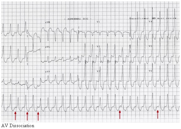
Ventricular tachycardia (VT) is usually a consequence of structural or ischemic heart disease, with breakdown of normal conduction patterns. Abnormal automaticity (which tends to favor ectopic foci) or activation of reentrant pathways in the myocardium can exist to generate the dysrhythmia. Electrolyte disturbances, ischemia, and sympathomimetics may increase the likelihood of VT in the susceptible myocardium.AV dissociation, shown in the image below, is apparent in approximately half of VT episodes, and when present, it is a hallmark characteristic of VT.1 This occurs because the sinus node is depolarizing the atria at a rate that is slower than the pathologic, faster ventricular rate. P waves can be visualized at times in between or embedded in the QRS complexes, but the P waves and QRS complexes have their own independent rates.
AV dissociation.
16 December 2010
THE NATURE AND EXPERIENCE OF PAIN
Pain is a universal human experience and the most common reason people seek medical care. Pain tells us something is wrong in the structure or function of our body and that we need to do something about it. Because pain is such a strong motivator for action, it is considered one of the body's most important protective mechanisms.Definitions of Pain
The International Association for the Study of Pain defined pain as "an unpleasant, subjective, sensory and emotional experience associated with actual or potential tissue damage or described in terms of such damage" (1979). Pain, however, is much more than a physical sensation caused by a single entity. It is subjective and highly individual, a complex mechanism with physical, emotional, and cognitive components.Pain cannot be objectively measured in the same way as, for example, the chemical content of urine or the oxygen content level of blood. Only the person who is suffering knows how the experience feels. For these reasons, McCaffery defined pain as "whatever the experiencing person says it is and whenever he says it does" (1979). The American Pain Society goes further by stating that it is "not the responsibility of clients to prove they are in pain; it is the nurse's responsibility to accept the client's report of pain" (2005).
Pain alters the quality of life more than any other health-related problem. It interferes with sleep, mobility, nutrition, thought, sexual activity, emotional well-being, creativity, and self-actualization. Surprisingly, even though pain is such an important obstacle to comfort, it is one of the least understood, most undertreated, and oft-discounted problems of healthcare providers and their clients. For this reason, some nurses and therapists add comfort to Maslow's hierarchy of basic human needs (1968). The American Pain Society goes further, declaring the relief of pain a "basic human right" (2005).
Traditionally, pain was considered merely a physical symptom of illness or injury, a simple stimulus-response mechanism. Though the historic role of nurses and therapists has been to relieve pain and suffering, there has been little understanding of the complexity of pain and only limited ways to manage it. Recent research shows pain to be a distinct disorder, with physical, emotional, and cognitive components. This view of pain has broadened our understanding of pain and given us new ways to understand its characteristics.
PAIN-RELATED TERMINOLOGY
Algesia: Sensitivity to pain.
Breakthrough pain: Transitory increase in pain to a level greater than the client's well-controlled baseline level (McCaffery & Pasero, 2003).
Hyperalgesia: Excessive sensitivity to pain.
Idiopathic pain: Chronic pain for which there is no identifiable psychological or physical cause.
Intractable pain: Pain that is not relieved by ordinary medical, surgical, and nursing measures (Mosby's Dictionary, 2009).
Pain threshold: Amount of pain required before individuals feel the pain. The lower the threshold, the less pain they can endure; the higher the threshold, the more pain they can endure.
Pain tolerance: Maximum amount and duration of pain a person can endure. Tolerance varies widely among people and is influenced by emotions and cultural background.
Pain syndrome: A group of symptoms of which pain is the critical element, such as headaches and post-herpetic neuralgia.
Phantom limb pain: Pain that occurs in a limb after it is removed or as a result of severe damage to the affected nerve plexus due to perceptual disruption in the brain.
Psychogenic pain: Chronic pain with no identified organic explanation.
Radiating pain: Pain that begins at one place and extends out into nearby tissues.
Referred pain: Pain that is felt at a different location than where tissue was damaged. This phenomenon occurs because pain fibers in the damaged area synapse near fibers from other areas of the body; for example, a myocardial infarction may create referred pain in the left shoulder.
Breakthrough pain: Transitory increase in pain to a level greater than the client's well-controlled baseline level (McCaffery & Pasero, 2003).
Hyperalgesia: Excessive sensitivity to pain.
Idiopathic pain: Chronic pain for which there is no identifiable psychological or physical cause.
Intractable pain: Pain that is not relieved by ordinary medical, surgical, and nursing measures (Mosby's Dictionary, 2009).
Pain threshold: Amount of pain required before individuals feel the pain. The lower the threshold, the less pain they can endure; the higher the threshold, the more pain they can endure.
Pain tolerance: Maximum amount and duration of pain a person can endure. Tolerance varies widely among people and is influenced by emotions and cultural background.
Pain syndrome: A group of symptoms of which pain is the critical element, such as headaches and post-herpetic neuralgia.
Phantom limb pain: Pain that occurs in a limb after it is removed or as a result of severe damage to the affected nerve plexus due to perceptual disruption in the brain.
Psychogenic pain: Chronic pain with no identified organic explanation.
Radiating pain: Pain that begins at one place and extends out into nearby tissues.
Referred pain: Pain that is felt at a different location than where tissue was damaged. This phenomenon occurs because pain fibers in the damaged area synapse near fibers from other areas of the body; for example, a myocardial infarction may create referred pain in the left shoulder.
Types of Pain
Pain is classified as acute and chronic. Acute pain has an identifiable cause and occurs soon after an injury to tissues in the body, such as bone, skin, or muscle. Acute pain is protective in that it motivates a person to take action. Its onset may be sudden or slow, and its intensity may vary from mild to severe. Acute pain is temporary and subsides as healing takes place. Severe acute pain activates the sympathetic nervous system, causing diaphoresis, increased respiratory and pulse rates, and elevated blood pressure.Chronic pain lasts beyond an expected healing phase, is non-protective in that it serves no function, and may not have an identifiable cause (Patterson, 2007). When pain goes on more than 6 months, it moves from being a "symptom" to a "condition." Chronic pain afflicts more than 1 in 5 Americans and is one of the most pervasive and thorny medical conditions in the United States. Such pain is described as nonmalignant (noncancerous), malignant (cancerous), and intractable.
- Nonmalignant, noncancerous chronic pain typically accompanies such conditions as osteoarthritis and peripheral neuropathy. The lack of purpose and uncertainty of duration of such pain may lead to depression, fatigue, insomnia, anorexia, apathy, learned helplessness, and other psychosocial consequences. If it is severe, chronic pain—like prolonged stress—activates the parasympathetic nervous system, resulting in muscle tension, decreased heart rate and blood pressure, and failure of body defenses.
- Malignant, cancerous chronic pain may be due to tumor progression, invasive procedures, infection, toxicities of treatment, and physical limitations. Such pain may be felt at the tumor site or some distance from it. Since clients with cancer may experience both chronic and acute pain, caregivers need to investigate immediately any new pain in these clients.
- Intractable chronic pain is pain from any cause that is resistant to medical, surgical, and nursing measures. Persistent and ongoing, intractable chronic pain may be psychogenic in nature (Mosby's Dictionary, 2009).
Sources of Pain
10 December 2010
Coronary artery disease is atherosclerosis of the coronary arteries that leads to a restriction of blood flow to the heart. Atherosclerosis is a chronic condition that narrows arteries by building fat-filled bulges in the arterial walls. These bulges are called atherosclerotic plaques, or simply, plaques. In some people, the plaques eventually break open, and the contents cause blood clots. If these clots are swept into the bloodstream, they can lodge in the smaller arteries downstream and completely block blood flow beyond that point.
Heart muscle is constantly active, and it requires a continuous blood supply. When a heart artery is blocked suddenly, the heart muscle it supplies stops working within a few minutes. If the blood supply remains blocked for a half-hour or more, the heart's muscle cells will begin to die.
Complete, sudden blockage of an artery is not the only problem. Even a reduced blood supply will reduce the oxygen supply to heart muscle, and oxygen-starved heart muscle responds with a characteristic feeling of pain or discomfort, called angina.
When its arteries are narrowed by atherosclerosis, a heart may still get enough oxygen to pump blood at rest. On the other hand, exercise increases the work of the heart, and narrowed arteries cannot always delivery the excess oxygen required by an exercising heart. Under these circumstances, a person with narrowed coronary arteries will get angina when exercising. One of the first symptoms of coronary artery disease is the appearance of angina when a person is working strenuously.
As atherosclerosis progresses, some of the plaques rupture and clots are formed. If a clot temporarily shuts down an artery, the patient will get sudden angina that lasts until the clot is broken or swept away. If the clot remains for an extended period of time, some heart muscle begins to die: this is a heart attack, or myocardial infarction.
In general, the sudden blockage of a coronary artery or one of its main branches is called an acute coronary syndrome, and myocardial infarctions are one form of acute coronary syndrome. Acute coronary syndrome is a medical emergency, and must be treated immediately in an emergency room.
Atherosclerosis, the cause of coronary artery disease, is a slow, long-term process. Typically, atherosclerosis begins in a person's teenage years or earlier, and the disease worsens quietly for decades. As people age, their atherosclerosis becomes more likely to involve the arteries of the heart and to become coronary artery disease.
The progression of atherosclerosis can be slowed or even stopped by a few preventive measures. These include stopping smoking, staying thin or losing weight, exercising regularly, and eating a low-fat balanced diet. To control atherosclerosis, it is also important to keep blood pressures low, to reduce high blood cholesterol levels, and to treat diabetes.
People who develop symptomatic coronary artery disease should begin or continue these anti-atherosclerotic programs. In addition, they should take aspirin daily, and they should probably take other medicines (typically, beta blockers) to reduce the workload of the heart. Nitroglycerin tablets can be used to alleviate occasional anginal pain, and surgical procedures are available to widen narrowed arteries.
Coronary artery disease is the number one killer in the developed world. In the United States, it is estimated that half of today's healthy 40-year-old men and a third of today's healthy 40-year-old women will eventually develop coronary artery disease.
The proportion of deaths in the United States that are due to coronary artery disease has been decreasing slowly but continuously over the past half-century. Nonetheless, coronary artery disease remains the single most common cause of death in the United States. One-fifth of all American deaths are attributed to coronary artery disease, and four-fifths of the deaths of people 65 years and older result from the disease.
Coronary artery disease is not just an American problem. Throughout the developed world, coronary artery disease causes more deaths and disabilities and is responsible for more economic costs than any other single illness. Moreover, it is predicted that, by the year 2020, coronary artery disease will have become the leading cause of death in the underdeveloped parts of the world.
The high death rates from coronary artery disease can be reduced by changing people's eating habits, by increasing their activity levels, and by convincing them of the dangers of cigarette smoking.

Heart muscle is constantly active, and it requires a continuous blood supply. When a heart artery is blocked suddenly, the heart muscle it supplies stops working within a few minutes. If the blood supply remains blocked for a half-hour or more, the heart's muscle cells will begin to die.
Complete, sudden blockage of an artery is not the only problem. Even a reduced blood supply will reduce the oxygen supply to heart muscle, and oxygen-starved heart muscle responds with a characteristic feeling of pain or discomfort, called angina.
When its arteries are narrowed by atherosclerosis, a heart may still get enough oxygen to pump blood at rest. On the other hand, exercise increases the work of the heart, and narrowed arteries cannot always delivery the excess oxygen required by an exercising heart. Under these circumstances, a person with narrowed coronary arteries will get angina when exercising. One of the first symptoms of coronary artery disease is the appearance of angina when a person is working strenuously.
As atherosclerosis progresses, some of the plaques rupture and clots are formed. If a clot temporarily shuts down an artery, the patient will get sudden angina that lasts until the clot is broken or swept away. If the clot remains for an extended period of time, some heart muscle begins to die: this is a heart attack, or myocardial infarction.
In general, the sudden blockage of a coronary artery or one of its main branches is called an acute coronary syndrome, and myocardial infarctions are one form of acute coronary syndrome. Acute coronary syndrome is a medical emergency, and must be treated immediately in an emergency room.
Atherosclerosis, the cause of coronary artery disease, is a slow, long-term process. Typically, atherosclerosis begins in a person's teenage years or earlier, and the disease worsens quietly for decades. As people age, their atherosclerosis becomes more likely to involve the arteries of the heart and to become coronary artery disease.
The progression of atherosclerosis can be slowed or even stopped by a few preventive measures. These include stopping smoking, staying thin or losing weight, exercising regularly, and eating a low-fat balanced diet. To control atherosclerosis, it is also important to keep blood pressures low, to reduce high blood cholesterol levels, and to treat diabetes.
People who develop symptomatic coronary artery disease should begin or continue these anti-atherosclerotic programs. In addition, they should take aspirin daily, and they should probably take other medicines (typically, beta blockers) to reduce the workload of the heart. Nitroglycerin tablets can be used to alleviate occasional anginal pain, and surgical procedures are available to widen narrowed arteries.
Coronary artery disease is the number one killer in the developed world. In the United States, it is estimated that half of today's healthy 40-year-old men and a third of today's healthy 40-year-old women will eventually develop coronary artery disease.
The proportion of deaths in the United States that are due to coronary artery disease has been decreasing slowly but continuously over the past half-century. Nonetheless, coronary artery disease remains the single most common cause of death in the United States. One-fifth of all American deaths are attributed to coronary artery disease, and four-fifths of the deaths of people 65 years and older result from the disease.
Coronary artery disease is not just an American problem. Throughout the developed world, coronary artery disease causes more deaths and disabilities and is responsible for more economic costs than any other single illness. Moreover, it is predicted that, by the year 2020, coronary artery disease will have become the leading cause of death in the underdeveloped parts of the world.
The high death rates from coronary artery disease can be reduced by changing people's eating habits, by increasing their activity levels, and by convincing them of the dangers of cigarette smoking.
TERMS RELATED TO CAD
Coronary artery disease (CAD) is the result of atherosclerosis of the coronary arteries of the heart. Coronary artery disease is also called:
- Coronary heart disease (CHD)
- Ischemic heart disease (IHD)
- Atherosclerotic heart disease
- Coronary atherosclerotic disease
- Chronic stable angina
- Acute coronary syndromes
- Unstable angina
- Myocardial infarction
- Sudden cardiac death
NORMAL CIRCULATION OF THE HEART
The heart is made up almost entirely of muscle. Cardiac muscle, which differs from the skeletal and smooth muscle of the rest of the body, is dependent on aerobic metabolism. This means that the heart cannot function without a constant supply of oxygen.A drawing of the heart, seen from the front. The coronary arteries and their main branches are large, and they run along the outer surface of the heart. The smaller arteries, which directly feed the heart muscle, dive deep into the walls of the heart (NHLBI, 2007c).
16 November 2010
To change proxy settings:
• Select Tools and then Internet Options.
• Click the Connections tab.
• If you are using a LAN, click the LAN Settings button. If you are using a Dial-up or Virtual Private Network connection, select the necessary connection and click the Settings button.
• Make sure the 'automatically detect proxy settings' and 'use a proxy automatic configuration script' options are not checked.
• In the 'Proxy Server' area, select the Use a proxy server for this connection or Use a proxy server for your LAN check box.
• In the Address box, enter the address of the proxy server. In the Port box, type the port number. If need to use different proxies for different services, such as HTTP, HTTPS, or FTP, click the Advanced button, and then enter the individual proxy server addresses to use.
• If necesary, enable the 'Bypass proxy server for local addresses' option.
• Click OK.
How to change Internet Explorer 7.x proxy settings
Subscribe to:
Posts (Atom)